Free Synapses revision notes for OCR A Level Biology – covering specification point 5.1.3 (d).
A synapse is the junction between a neurone and a postsynaptic cell (usually another neurone), over which action potentials cannot propagate.
The diagram below shows the synaptic cleft between the presynaptic bulb and the opposing dendritic membrane of a postsynaptic neurone:
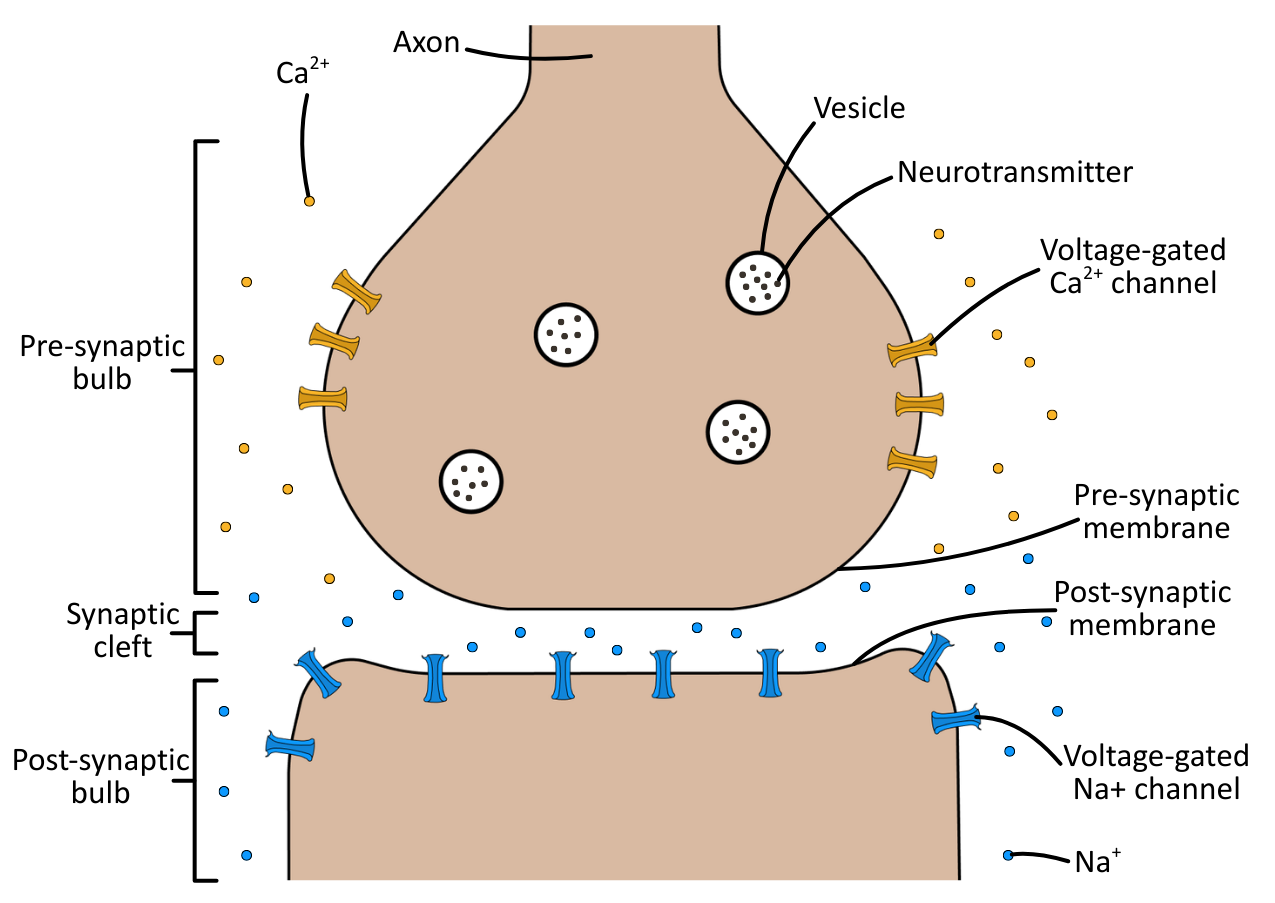
Neurones communicate across this gap chemically, releasing neurotransmitters from the presynaptic membrane that diffuse across the synaptic cleft, and bind to receptors on the postsynaptic membrane.
Types Of Synapse
There are two main types of synapse, inhibitory and excitatory:
- Inhibitory synapses hyperpolarise postsynaptic membranes, increasing the potential difference away from the threshold potential and making an action potential less likely.
- Excitatory synapses depolarise postsynaptic membranes, decreasing the potential difference towards the threshold potential and making action potentials more likely.
Additionally, synapses use different neurotransmitters, depending on their function. For example, cholinergic synapses use acetylcholine, dopaminergic synapses use dopamine, and GABAergic uses γ-aminobutyric acid. In OCR A Level Biology cholinergic synapses are the named example you are required to know.
Summation
Summation is the accumulative effect of postsynaptic potentials at the postsynaptic membrane, which, when combined, can generate an action potential if they reach the threshold potential.
It is important to note that the postsynaptic potentials can be either excitatory or inhibitory:
- Excitatory: A small depolarisation of the postsynaptic membrane, known as an EPSP (excitatory postsynaptic potential).
- Inhibitory: A small hyperpolarisation of the postsynaptic membrane, known as an IPSP (inhibitory postsynaptic potential).
The EPSPs and IPSPs combine (so can cancel each other out), and the firing of an action potential is dependent upon the threshold potential being reached.
How Synapses Work
Presynaptic neurones release neurotransmitters stored in vesicles into the synaptic cleft via exocytosis, which then diffuse across to the postsynaptic membrane.
Postsynaptic membranes have specific receptors to which neurotransmitters can bind, that open their ion channels, resulting in an excitatory (depolarising) or inhibitory (hyperpolarising) effect on the postsynaptic membrane.
The diagram below shows the mechanism of excitatory synaptic action:
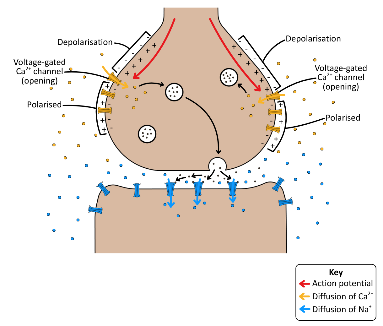
Neurotransmitter in the synaptic cleft is either broken down by enzymes in the synaptic cleft or reabsorbed by the presynaptic bulb, which ensures that the transmission of an impulse from one neurone to the next is brief and discrete.
Want to dive deeper?
-

Textbook: Introduction to Synapses
£1.99 -

Textbook: Synaptic Action
£2.99 -

Bundle: Year 12 Full Access
£79.99 -

Bundle: Year 12 Textbook
£44.99