Free Kidney Anatomy and Histology revision notes for OCR A Level Biology – covering specification point 5.1.2 (cii).
The Kidney
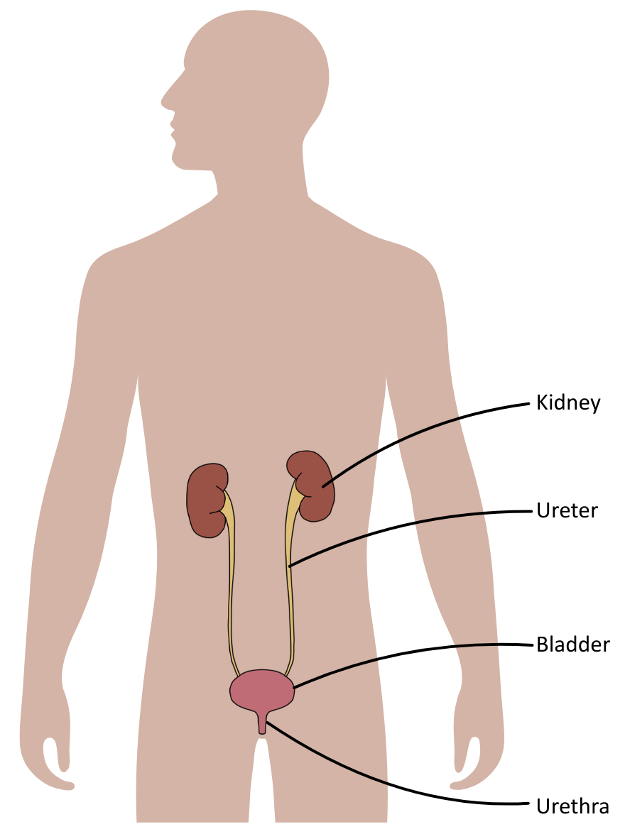
Humans have two kidneys, located on either side of the vertebral column, just below the lowest rib.
The diagram below shows the position of the kidneys:
Kidneys are the main organs for excretion and osmoregulation, and play a central role in homeostatic processes that maintain the optimal composition of the blood plasma and tissue fluid, including:
- Excretion: Removal of metabolic wastes such as urea and creatinine from the blood.
- Water balance: Regulation of blood water potential through the controlled reabsorption of water and production of urine.
Anatomy of the Kidney(s)
The kidney has 3 tissue areas arranged around a central region (the renal pelvis), which collects urine before it enters the ureter. Surrounding the renal pelvis is the medulla, and surrounding that is the cortex.
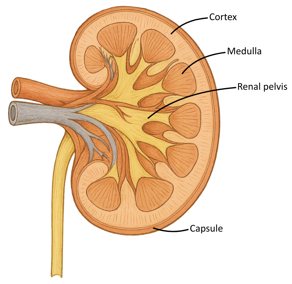
The table below outlines the regions of the kidney and their role(s):
| Region | Description | Main Function |
|---|---|---|
| Cortex | The outer region contains renal corpuscles and convoluted tubules. | Site of ultrafiltration and the selective reabsorption of most useful solutes. |
| Medulla | The inner region is composed of renal pyramids, containing loops of Henle and collecting ducts. | Establishes an osmotic gradient to enable water reabsorption. |
| Renal pelvis | The central region where the collecting ducts drain into the ureter. | Funnels urine to the bladder. |
Histology of the Kidney(s)
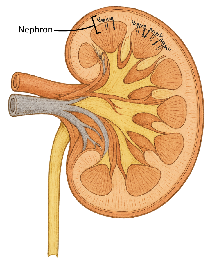
A nephron is a tubular structure responsible for filtering the blood and forming urine.
The nephron is a functional unit of the kidney, with approximately 1 million of them arranged radially across the cortex and medulla, converging toward the pelvis.
The diagrams below show the position of a single and many nephrons in the kidney:
It is important to note that the collecting duct is not a component of the nephron; however, in A level materials, it is usually referred to as such.


