Free Neuronal Control of Striated Muscle OCR A Level Biology revision notes – covering specification points 5.1.5 (li) and 5.1.5 (lii).
Want to go deeper?
Neuronal Control of Striated Muscle: Neuromuscular Junctions
Skeletal muscle contracts only when stimulated by a motor neurone.
The specialised synapse between a motor neurone and a muscle fibre is known as a neuromuscular junction.
The region of the muscle fibre membrane (the sarcolemma) that faces the synaptic terminal is known as the muscle end plate.
A neuromuscular junction’s mechanism of action can be summarised as:
- An action potential arrives at the synaptic terminal.
- Voltage-gated Ca²⁺ ion channels open, allowing Ca²⁺ ions to diffuse in down their concentration gradient.
- Ca²⁺ ions cause vesicles containing acetylcholine to undergo exocytosis.
- Acetylcholine diffuses across the synaptic gap and binds to receptors on the sarcolemma.
- Na⁺ channels open, allowing Na⁺ to diffuse into the muscle fibre, depolarising it.
- Ca²⁺ ions are released from the sarcoplasmic reticulum, initiating muscle contraction.
Acetylcholine is quickly broken down by acetylcholinesterase to prevent continuous stimulation of the neuromuscular junction and avoid unneeded contractions.
Muscle Contraction: Myofibrils and the Sliding Filament Theory
The sliding filament theory (also known as the sliding filament model, or hypothesis) is the mechanism by which skeletal muscle fibres contract.
Sarcomeres are the contractile elements of a muscle fibre, consisting of the proteins actin and myosin.
The diagram below shows the overlapping structure of actin and myosin:
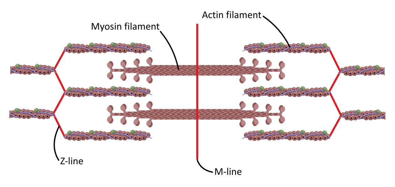
During contraction, the actin and myosin filaments overlap, which changes the size of the light and dark regions.
The table below outlines the features which can be observed in skeletal muscle tissue under an optical microscope and their significance:
| Feature | Appearance | Significance |
|---|---|---|
| A-band | Dark band | Thick myosin filaments, which can overlap with actin |
| H-zone | Lighter region in the centre of the A-band | Thick filaments only |
| I-band | Light band | Thin filaments only |
| Z-line | Thin dark line | Sarcomere boundary |
The Sliding Filament Model
The process can be summarised as:
- Ca²⁺ ions are released from the sarcoplasmic reticulum and bind to troponin.
- Troponin’s conformational shape changes, moving tropomyosin away from the myosin-binding sites on actin and exposing them.
- Myosin heads bind to the myosin-binding site on actin, forming a cross-bridge.
- Myosin heads move by tilting, sliding the thin filament along the myosin filament, and releasing ADP and an inorganic phosphate (Pi).*
- ATP binds to the myosin head, causing the cross-bridge to break and the myosin head to detach from actin.
- ATP is then hydrolysed by myosin ATPase, providing the energy to return the myosin head to its high-energy ‘cocked’ position**.
*This is known as the powerstroke.
**Described in some sources as re-cocking.
Creatine phosphate is a store of phosphate groups in the sarcoplasm that can be used to regenerate ATP quickly for use in sarcomere contraction.