Free Mammalian Muscle Structure OCR A Level Biology revision notes – covering specification point 5.1.5 (li).
Want to go deeper?
Mammalian Muscle
Muscles are effectors that produce movement responses in animals.
The 3 main types of muscle in mammals are cardiac, skeletal and smooth:
- Skeletal muscle* is connected to the skeleton by tendons, contracting in response to conscious stimulation from the somatic nervous system.
- Cardiac muscle* is found in the heart and contracts rhythmically without becoming fatigued.
- Smooth muscle is found in tubular structures, contracting slowly and rhythmically in response to unconscious stimulation from the autonomic nervous system.
*Both cardiac and skeletal muscle are types of striated muscle (skeletal often being incorrectly treated as synonymous with striated muscle). Cardiac muscle is often (and misleadingly) treated as its own separate type at A level for simplicity, as we have done here.
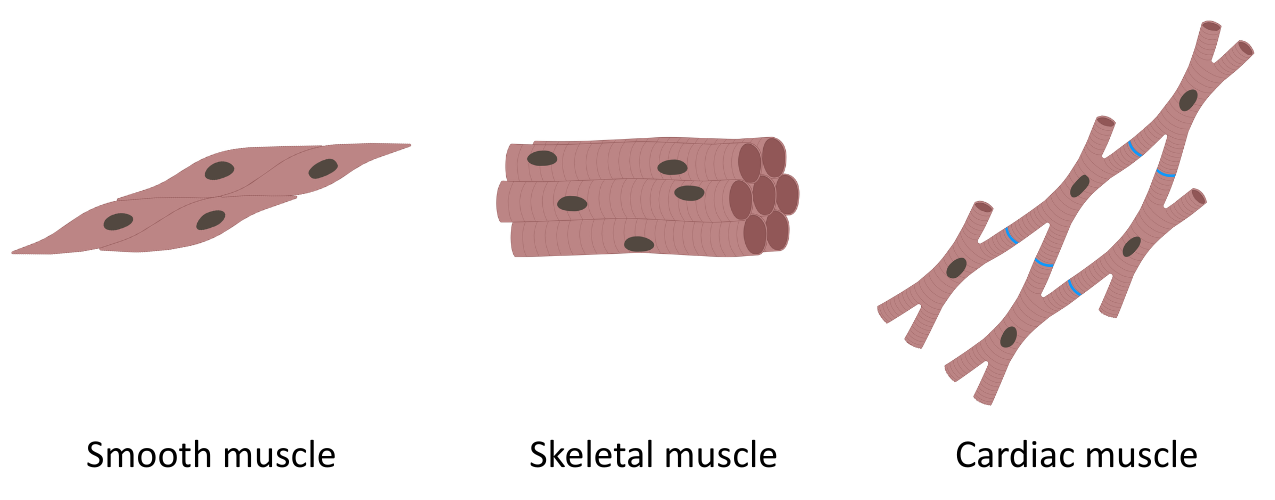
The diagram below shows the typical appearance of skeletal, cardiac and smooth muscle:
Skeletal Muscle
A skeletal muscle fibre is a highly specialised cell formed from the fusion of several cells to form an elongated cell with many nuclei along its length.
The cell surface membrane of a skeletal muscle cell is known as a sarcolemma.
The sarcolemma is a specialised cell surface membrane with several features adapted to its role in muscle contraction:
- Motor end plates: Regions of the membrane facing the axon terminals of motor neurones, with acetylcholine receptors that can initiate depolarisation of the sarcolemma.
- T-tubules: The sarcolemma folds inwards, forming deep transverse tubules (T-tubules) that can propagate an action potential deep into a muscle fibre, allowing the entire fibre to contract (almost) simultaneously.
The cytoplasm of a skeletal muscle cell is known as the sarcoplasm, and is highly specialised:
- Many myofibrils: Contractile elements made up of the proteins actin and myosin.
- Many mitochondria: Perform aerobic respiration to make lots of ATP for contraction.
- Extensive sarcoplasmic reticulum: A highly specialised smooth endoplasmic reticulum wrapped around the myofibrils, which releases stored Ca²⁺ in response to neuronal stimulation of the muscle fibre.
Cardiac Muscle
The structure of a cardiac muscle cell is that of a short cell with branches to join with other cardiac muscle cells. It usually has one nucleus, but sometimes has two.
It is important to note that cardiac muscle cells have many similarities with skeletal muscle cells, and that many details set out in the OCR A Level Biology endorsed textbook relating to skeletal muscles are also true of cardiac muscle.
Smooth Muscle
Smooth muscle is a type of involuntary muscle found mainly in the walls of tubular organs (such as blood vessels, the digestive system, uterus), where it can control the movement of substances through the body.
Smooth muscle contracts slowly, but can maintain its state of contraction for a long time, making it resistant to fatigue.
It is important to note that you are not expected to know the structure of a smooth muscle cell.
Comparing Types of Mammalian Muscle
The table below compares some of the key features of each type of muscle cell:
| Feature | Skeletal Muscle | Cardiac Muscle | Smooth Muscle |
|---|---|---|---|
| Control | Voluntary | Involuntary | Involuntary |
| Cell shape | Long, cylindrical fibres | Short, branching cells | Spindle-shaped |
| Striations | Present (has sarcomeres) | Present (has sarcomeres) | Absent (no sarcomeres) |
| Intercalated discs | Not present | Present | Not present |
| Presence of sarcomeres | Yes | Yes | No |