Module 4: White Blood Cells and Secondary Defences
These free OCR A Level Biology White Blood Cells and Secondary Defences revision notes have been written for specification points 4.1.1(e.i), 4.1.1(e.ii) and 4.1.1(f).
White Blood Cells
Different roles in the immune response are performed by different types of white blood cells, each specialised for their role.
The table below lists the different types of white blood cells named in the OCR A level Biology course, and what type they are:
| White Blood Cell | Phagocyte | Antigen-Presenting Cell (APC) | Lymphocyte | Non-specific Response | Specific Immune Response |
|---|---|---|---|---|---|
| Neutrophil |  | | |||
| Monocyte / Macrophage | | | | ||
| Plasma Cell / B effector cell | | | |||
| B Memory | | | |||
| T Helper | | | |||
| T Killer | | | |||
| T Memory | | | |||
| T Regulatory | | |
Identifying white blood cells
The main types of white blood cells you need to know can be identified from a blood smear under a microscope:
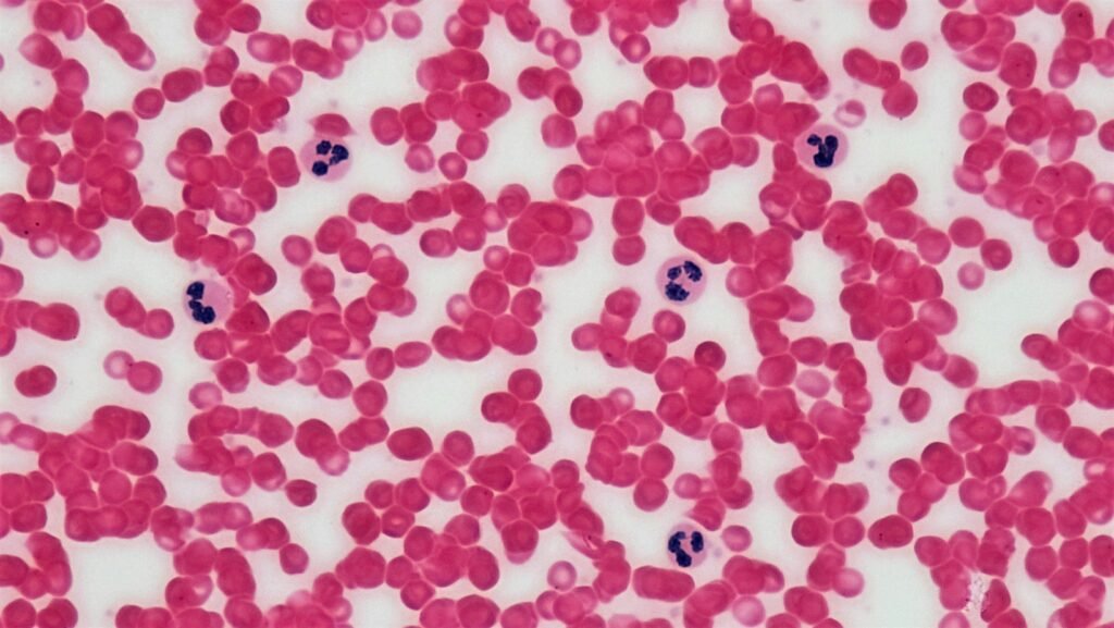
The table below outlines the main distinguishing features you would be expected to recognise in an exam:
| Lymphocyte | Distinguishing Feature | Image |
|---|---|---|
| Neutrophils | Multi-lobed nucleus |  |
| Lymphocytes | Large round nucleus |  |
| Monocytes / Macrophages | Kidney-shaped nucleus Larger than other lymphocytes |  |
Secondary Defences: Non-Specific and the Specific Immune Response
Secondary defences are for when pathogens get past primary defences and enter the body.
The secondary defences of the immune system are either:
- Non-specific: The response of white blood cells is indiscriminate, targeting any ‘foreign’ antigens.
- Specific: The response of white blood cells is controlled by the presence of particular antigens which they target.
Macrophages are non-specific in how they approach phagocytosis, but their display of antigens involves them in the start of the specific immune response.
Only phagocytes are involved in the secondary non-specific immune response.
Phagocytes
Phagocytes are cells that do phagocytosis: the engulfing of a pathogen and destroying it with hydrolytic enzymes.
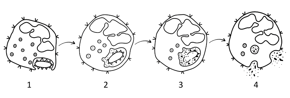
The process of phagocytosis is:
- Recognition: The phagocyte binds to the antigen on the pathogen’s surface.
- Engulfment: The pathogen is engulfed by endocytosis and enclosed in a phagosome.
- Digestion: Lysosomes fuse with the phagosome and release hydrolytic enzymes.
- Absorption or Release: Harmless products are absorbed or expelled to be used as nutrients.
The two types of phagocyte taught in OCR A level Biology are neutrophils and macrophages.
The table below compares the main features of neutrophils and macrophages that you need to know:
| Feature | Neutrophils | Macrophages |
|---|---|---|
| Origin | Bone marrow | Bone marrow (as monocytes) |
| Maturation site | Bone marrow | Lymph nodes (and other tissues) (differentiating from monocytes) |
| Lifespan | Short-lived (hours to days) | Long-lived (can persist for months) |
| Role | Phagocytosis of pathogens |
– Phagocytosis – Antigen presentation |
| Structure |
– Multi-lobed nucleus – Many lysosomes – Many mitochondria – Many ribosomes – Chemotaxis |
– Kidney-shaped nucleus* – Many lysosomes – Many mitochondria – Many ribosomes – Chemotaxis |
*Monocytes have the kidney shaped nucleus, which becomes irregular as they mature into macrophages.
Non-specific immune response
In the non-specific immune response neutrophils and macrophages carry out phagocytosis on any foreign microorganisms they find.
Microorganisms are recognised as being foreign due to the antigens they have.
Neutrophils and macrophages are ‘non-specific’ because they will respond to any antigen, not just certain ones (as happens in the specific immune response).
The specific immune response
The specific immune response identifies certain pathogens by recognising their antigen marker molecules and targeting them (mostly) exclusively.
This allows pathogens, or infected cells, to be destroyed more effectively. The immune system can then make powerful antibodies that inhibit the pathogen’s function. It also provides immunity by recognising the same pathogen upon reinfection.
The main types of white blood cells involved are:
- B cells: Responsible for making antibodies that target specific antigens.
- T cells: Responsible for attacking infected host cells and stimulating B cells.
Both B and T lymphocytes are involved in immunity by making memory cells.
The actions of B and T lymphocytes in the specific immune response are driven by 4 processes:
- Antigen presentation: Where a cell displays an antigen on its cell surface membrane to activate specific B and T cells with a complementary receptor.
- Clonal selection: Where specific T and B lymphocytes are activated after binding with a complementary antigen on an antigen-presenting cell (APC).
- Clonal expansion: Where activated B and T lymphocytes divide by mitosis; each new cell is able to produce complementary receptors or antibodies to the same antigen.
- Differentiation: Where some B and T cells are produced by clonal selection and turn into specialised lymphocytes.
The activity of the immune system is controlled by cell signalling, including:
- Antigen-presentation: Some lymphocytes have complementary protein receptors to specific antigens, and binding to these, either directly on pathogens or on APCs, will activate them.
- Cell signalling molecules: Chemical messengers between B and T cells stimulate the immune system (e.g. cytokines and interleukins*), whilst other body cells can release substances to alert lymphocytes that they are infected (e.g. interferon for viral infections).
*Technically, interleukins are just a type of cytokine, but this is misrepresented in many educational materials.
B and T Cells
The table below outlines the roles of the lymphocytes involved in specific immunity:
| Cell Type | Function |
|---|---|
| Undifferentiated lymphocyte |
– Detects antigens using complementary receptors on its surface. – Activated via antigen presentation and clonal selection, then undergoes clonal expansion. |
| B Memory cell |
– Provides long-term immunity. – Responds rapidly to known antigens by undergoing clonal expansion. |
| Plasma Cell / B Effector Cell | – Produces and secretes antibodies specific to one antigen. |
| T Memory Cell |
– Provides long-term immunity. – Responds rapidly to known antigens by undergoing clonal expansion. |
| T Killer Cell |
– Destroys infected cells that activate it with antigen presentation and interleukins. – Releases perforins and hydrolytic enzymes. |
| T Helper Cell |
– Coordinates the immune response. – Releases interleukins to stimulate clonal selection and clonal expansion of B and T cells. |