Module 2: Biological Membranes
These free OCR A Level Biology Biological Membranes revision notes have been written for specification points 2.1.5(a), 2.1.5(b), 2.1.5(c), 2.1.5(d.i), 2.1.5(e.i) and 2.1.5(f).
Biological Membranes
Biological membranes are selectively (partially) permeable lipid barriers that enable the separation of a cell’s contents from its external environment.
In addition to controlling the movement of substances, membranes have many more functions important to both prokaryotic and eukaryotic cells, detailed in the table below:
| Role of Membrane | Structure(s) Involved | Main example |
|---|---|---|
| Control the entry and exit of substances |
– Phospholipid bilayer* – Proteins |
– Cell surface membrane – Organelle membranes (e.g. mitochondria, nucleus) |
| Cell communication |
– Glycoproteins – Receptor proteins – Vesicles | – Cell surface membrane |
| Cell recognition |
– Glycoproteins – Glycolipids | – Cell surface membrane |
| Chemical reactions | – Embedded enzymes |
– Inner mitochondrial membrane (aerobic respiration) – Thylakoid membranes (photosynthesis) |
| Maintains electrochemical gradients |
– Proton pumps – Ion channels |
– Inner mitochondrial membrane – Thylakoid membranes – Cell surface membrane |
| Transport and secretion | – Vesicles |
– Golgi apparatus – Endoplasmic reticulum – Cell surface membrane |
The Fluid Mosaic Model
The cell surface membrane consists of a phospholipid bilayer with proteins (and some other molecules) embedded in it.
At GCSE it was enough to call it ‘the cell membrane’, but at A level this is too vague to score any marks.
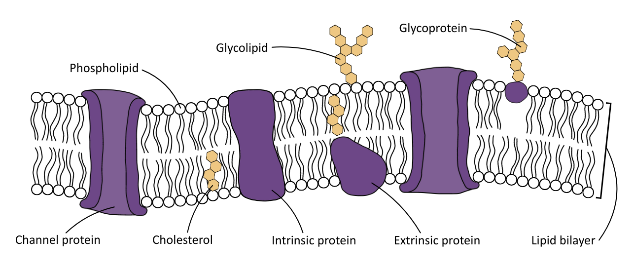
The protein components (e.g. glycoprotein, carrier protein) can be classified as either
- Integral proteins: Go from one side of the lipid bilayer to another
- Peripheral proteins: Are located only on one side of the lipid bilayer
Because the components are free to move around each other (it’s fluid) and the components are interspersed with each other (like a mosaic), this model of how the plasma membrane works is called the fluid mosaic model.
The table below outlines the components of the plasma membrane:
| Component | Structure | Function |
|---|---|---|
| Phospholipid bilayer | Two layers of phospholipids with hydrophobic fatty acid tails facing inwards and hydrophilic phosphate heads facing outwards |
– Provides a barrier to most water-soluble substances – Allows lipid-soluble molecules to pass – Allows small uncharged molecules to pass through |
| Cholesterol | Found between phospholipids |
– Gives mechanical stability and flexibility – Stabilises the membranes’ fluidity by reducing fluidity at high temperatures and preventing rigidity at low temperatures |
| Glycolipids | Phospholipids with a carbohydrate chain attached |
– Used in cell signalling and recognition – Stabilises the plasma membrane, as carbohydrate chains interact with the aqueous environment |
| Glycoproteins | Proteins with carbohydrate chains |
– Antigens – Receptors – Important in signalling and immune response – Stabilises the plasma membrane, as carbohydrate chains interact with the aqueous environment |
| Channel proteins | Globular proteins with a pore (integral) | Passive movement (diffusion) of ions and small polar molecules. |
| Carrier proteins | Globular proteins with a pore (integral) | Used in facilitated diffusion and active transport. |
| Embedded proteins | Globular proteins (peripheral) |
– Enzymes – Antigens – Receptors |
Membrane Permeability
The relative abundance of each component in a plasma membrane affects its permeability to different substances, for example:
| An Increase In… | Effect on Permeability |
|---|---|
| Phospholipids | ↑ permeability to small, non-polar molecules (e.g. O₂, CO₂) |
| Cholesterol | ↓ permeability to water and small polar molecules |
| Channel proteins | ↑ permeability to ions (e.g. Na⁺, K⁺, Cl⁻) |
| Carrier proteins | ↑ permeability to larger polar molecules (e.g. glucose) |
| Aquaporins | ↑ permeability to water |
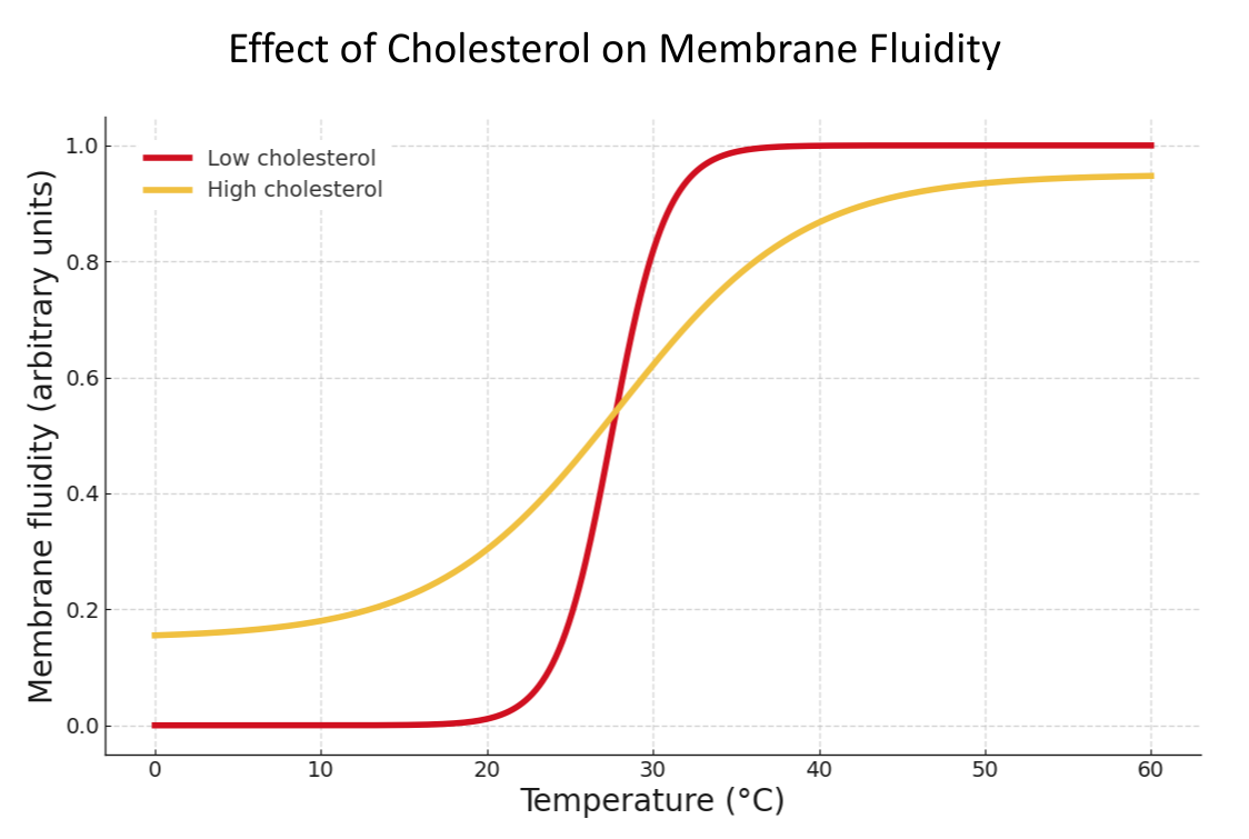
The structure of a membrane can be affected by environmental conditions listed in the table below:
| Factor | Effect | Mechanism |
|---|---|---|
| Low temperature |
– Membrane becomes less fluid and more rigid (brittle) – Permeability decreases | Saturated fatty acid tails on the phospholipids pack together more closely. |
| High temperature |
– The membrane becomes more fluid – Permeability increases – Proteins may denature |
– Phospholipids move more, so there are more gaps in the membrane – Tertiary structure bonding (hydrogen and ionic) disrupted or denatured |
| Solvents (e.g. ethanol) |
– Disrupt membrane structure – Increase permeability | Organic solvents dissolve lipids, disrupting the bilayer and allowing substances to leak through |
| pH changes | – Denatures membrane proteins | Alters ionic and hydrogen bonding in the tertiary structure |
| Detergents | – Break apart the membrane completely | Detergents emulsify phospholipids, disrupting the plasma bilayer |
The diagram below shows the combined effects of temperature and cholesterol on the fluidity of a plasma membrane.
Transport Across Membranes
Cellular transport processes are divided into two types:
- Active: Uses ATP
- Passive: Does not use ATP
The movement of substances in passive transport processes is driven by concentration gradients, from a high concentration to a low concentration.
The table below outlines the different transport processes:
| Process | Definition | Needs ATP? | Suitable molecules |
|---|---|---|---|
| Simple diffusion | Net movement from high to low concentration through the bilayer. |  | Small Non-polar Lipid soluble |
| Facilitated diffusion | Movement down conc. gradient via channel or carrier proteins. | | Small Polar Lipid insoluble |
| Osmosis | Net movement of water from high to low water potential across a plasma membrane. | | Water (only) |
| Active transport | Movement against a concentration gradient using ATP and carrier proteins. |  | Charged ions Polar molecules Lipid insoluble |
| Co-transport | Movement of one substance down its gradient pulls another against its gradient (ATP indirectly). | (indirect) | Small Polar Lipid insoluble |
| Endocytosis | Bulk transport into the cell via vesicle. | | Too large |
| Exocytosis | Bulk transport out of the cell via vesicle. | | Too large |
Effect of distance
Diffusion distance (mostly) applies to simple diffusion. It is just the idea that the further a substance has to move to get from ‘A to B’, the lower its rate of diffusion.
This is minimised in exchange surfaces to decrease the distance between ‘A and B’ as much as possible.
The table below gives the specialised exchange surfaces that minimise diffusion distance encountered in A level Biology:
| Exchange Surface | Adaptation | Substances |
|---|---|---|
| Alveoli |
– One-cell-thick alveolar wall – One-cell-thick capillary wall – Squamous epithelium | O₂, CO₂ |
| Capillaries | – One cell-thick endothelium | O₂, CO₂, glucose, amino acids |
| Villi and microvilli | – Single-layer epithelial cells | Glucose, amino acids, fatty acids |
| Root hair cells | – Thin cell wall | Water, mineral ions (e.g. nitrates) |
| Leaf mesophyll |
– Thin, flat cells – Air spaces between cells | CO₂, O₂ |
| Placenta | – A thin membrane between maternal and fetal blood | O₂, glucose, urea, CO₂ |
Effect of size (of molecule)
Smaller molecules diffuse at a faster rate than larger ones, which (mostly) applies to simple diffusion.
For processes using transport proteins, size mainly relates to whether or not the molecule can fit into the transport protein shaped specifically for it, and is irrelevant for bulk transport.
Effect of surface area (of the cell)
The greater the surface area, the more of a substance can cross the plasma membrane, at the same time, through its transport process.
In cells using transport proteins, the surface area may directly affect how many they have to use.
Specialised cells will have adaptations to increase their surface area.
The table below gives an overview of two specialised exchange surfaces that minimise diffusion distance:
| Exchange Surface | Adaptation to Increase Surface Area | Substances Exchanged |
|---|---|---|
| Alveoli |
– Millions of small alveoli – Folded internal structure | O₂, CO₂ |
| Root hair cells |
– Long, thin root hair extensions – Numerous root hairs | Water, mineral ions (e.g. nitrates) |