Module 3: The Heart
These free OCR A Level Biology The Heart revision notes have been written for specification points 3.1.2(e.), 3.1.2.(f), 3.1.2(g) and 3.1.2(h).
External Structure Of The Heart
The heart is a muscular organ located in the thoracic cavity between the lungs. It is enclosed in a tough, fluid-filled sac called the pericardium, which protects the heart and reduces friction as it beats.
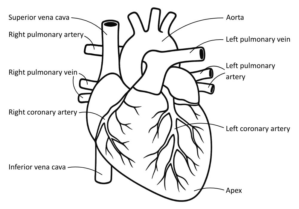
The table below outlines the heart’s external anatomical features:
| Feature | Description | Function |
|---|---|---|
| Cardiac muscle | The heart wall is made of myogenic striated muscle. | Contracts rhythmically and does not fatigue. |
| Coronary arteries | Blood vessels on the external surface that branch off from the aorta. | Supply oxygenated blood to the heart muscle. |
| Apex | The pointed lower end of the heart tilted towards the left. | Helps identify the left side of the heart in dissection or imaging. |
The following major blood vessels are visible on the outside of the heart:
| Blood Vessel | Description | Function |
|---|---|---|
| Vena cava | Large vein entering the right atrium from above (superior) and below (inferior). | Returns deoxygenated blood from the body. |
| Pulmonary artery | Emerges from the right ventricle; divides into two branches. | Carries deoxygenated blood to the lungs. |
| Pulmonary veins | Two veins from each lung enter the left atrium. | Return oxygenated blood from the lungs. |
| Aorta | Large artery leaving the left ventricle, arching over the heart. | Carries oxygenated blood to the rest of the body. |
Internal Structure Of The Heart
The mammalian heart is a double pump, keeping oxygenated and deoxygenated blood separated to ensure efficient oxygen transport.
The path blood takes through the heart, when returning deoxygenated blood from the body, is as follows:
[Systemic circuit] → Vena cava → Right atrium → Atrioventricular valve → Right ventricle → Semilunar valve → Pulmonary artery → [Pulmonary circuit] → Pulmonary veins → Left atrium → Atrioventricular valve → Left ventricle → Semilunar valve → Aorta → [Systemic circuit]
The table below outlines the structural components of the heart:
| Feature | Description | Function |
|---|---|---|
| Left atrium | Upper chamber on the left side. | Receives oxygenated blood from the lungs. |
| Right atrium | Upper chamber on the right side. | Receives deoxygenated blood from the body. |
| Left ventricle | Lower chamber on the left side; has a thicker muscular wall. | Pumps oxygenated blood to the body via the aorta. Must pump more strongly to transport blood further. |
| Right ventricle | Lower chamber on the right side; has a thinner muscular wall compared to the left chamber. | Pumps deoxygenated blood to the lungs via the pulmonary artery. |
| Septum | Muscular wall separating the left and right sides of the heart. | Prevents mixing of oxygenated and deoxygenated blood. |
| Atrioventricular (AV) valves | Membranes attached by elastic tissue found between the atria and ventricle. | Prevent backflow of blood from ventricles to the atria. |
| Semilunar valves | Membranes attached by elastic tissue found at the ventricle exits. | Prevent backflow of blood from arteries into the ventricles. |
| Coronary arteries | Branches from the aorta that return to the outside of the heart, supplying it with oxygenated blood. | Ensure the heart muscle gets oxygen and glucose for continuous aerobic respiration. |
The Cardiac Cycle
The cardiac cycle is the sequence of events that occurs during one complete heartbeat.
The table below outlines the 3 stages of the cardiac cycle:
| Stage | Key Events |
|---|---|
| Atrial systole |
Both atria contract, increasing the pressure in the atria. Blood is pushed through the AV valves into the ventricles. Ventricles remain relaxed. |
| Ventricular systole |
Both atria relax. Both ventricles contract, increasing the pressure in the ventricles. AV valves close, preventing backflow. Semilunar valves open, forcing blood into the pulmonary artery and the aorta. |
| Diastole |
Ventricles relax, and as the pressure drops, the semilunar valves close to prevent backflow from the arteries. Atrial pressure increases as blood flows passively into the atria. |
The table below compares the overall pressure and volume of blood in the heart across the different stages:
| Stage | Event | Pressure | Volume (of blood) | Notes |
|---|---|---|---|---|
| Diastole | Heart relaxes; chambers fill | ↓ | ↑ | AV valves open, SL valves closed |
| Atrial systole | Atria contract → ventricles fill | ↑ | Same | AV valves remain open |
| Ventricular systole | Ventricles contract → blood is forced out | ↑↑ | ↓ | AV valves shut, SL valves open |
Coordination Of Heart Contraction
The heart is myogenic, meaning it generates its own electrical impulses to control the rhythm of atrial and ventricular contraction without stimulation from the nervous system.
Electrical stimulation coordinates the atria and ventricular contractions, ensuring they do so in the right order and at the right time, to effectively move blood throughout the heart and prevent backflow.
Myogenic control of the heart is carried out by the components outlined in the table below:
| Component | Location & Structure | Function |
|---|---|---|
| Sinoatrial Node (SAN) | Right atrium wall. | Generates electrical impulses at regular intervals, acting as a natural pacemaker. |
| Atrioventricular Node (AVN) | In the upper septum between the atria and ventricles. | Delays the impulse slightly to allow the atria to finish contracting before the ventricles contract. |
| Bundle of His | Conductive fibres running down the septum wall to the apex of the heart. | Transmits impulses from the AVN to the Purkinje* fibres in the ventricles. |
| Purkinje fibres* | Spread through the ventricular walls up from the apex. | Distribute the impulse throughout the ventricular walls to ensure even ventricular contraction up from the apex. |
*Purkinje fibers are also known as Purkyne tissue.
The order of events is as follows:
SAN fires → atria contract → AVN delays the impulse → Bundle of His carries the impulse to Purkyne fibres → Purkyne fibres spread the impulse → ventricles contract from apex upwards
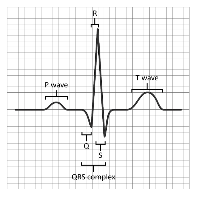
Electrocardiograms
The electrical activity of these events can be recorded, measured and observed with an ECG.
There are three distinct ‘waves’ of polarisation and depolarisation that can be observed:
- P wave: Depolarisation of the atria (they are electrically stimulated and contract).
- QRS complex: Depolarisation of the ventricles (they are electrically stimulated and contract).
- T wave: Repolarisation of the ventricles (the ventricles relax).
Heart conditions can be identified and classified with the use of ECG:
| Condition | ECG Feature | Trace | Cause |
|---|---|---|---|
| Tachycardia | Rapid heart rate: >100 bpm | 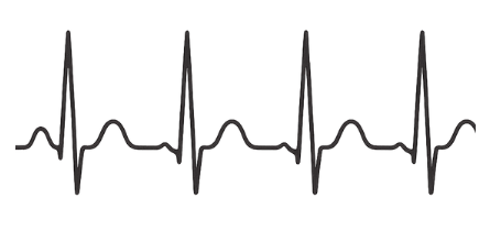 | Stress Fever Exercise |
| Bradycardia | Slow heart rate: <60 bpm | 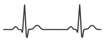 | Can be normal Disease |
| Ectopic beat | Early contraction of atria or ventricles. | 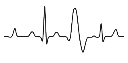 | Often harmless Can indicate arrhythmia. |
| Fibrillation | Uncoordinated contractions (an irregular trace). | 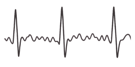 | Damage to myogenic structures |